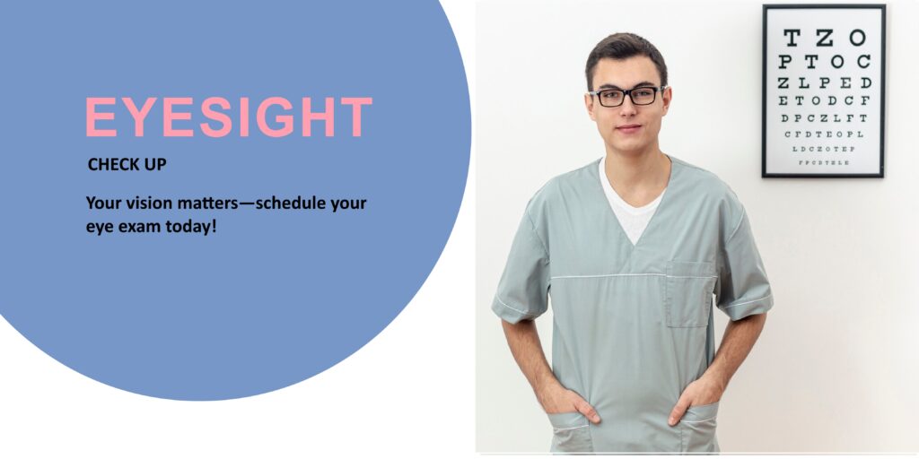
We are always ready to help you. Book an appointment
For Emergencies Contact: +91 8847270023

Detect vision problems early (e.g., nearsightedness, farsightedness, astigmatism)
Screen for eye diseases like glaucoma, cataracts, macular degeneration, and diabetic retinopathy
Monitor eye health changes if you already wear glasses or contact lenses
Identify signs of other health issues (e.g., diabetes, hypertension) that can show symptoms in the eyes
Children: First check at 6 months, then around 3 years, before starting school, and regularly thereafter
Adults (18–60 years): Every 2 years if no symptoms or vision problems; every year if you wear glasses or contacts
Adults (60+ years): Every year to monitor for age-related eye conditions
If you have risk factors (e.g., diabetes, family history of eye disease): Every year or as recommended by your doctor
Review of medical and vision history
Tests for visual acuity (sharpness of vision)
Eye pressure test (to check for glaucoma)
Refraction assessment (to update prescription if needed)
Eye health examination with special instruments (sometimes including pupil dilation)
You will be asked to read letters from an eye chart (commonly the Snellen chart) at a distance.
Distance vision is checked first.
Sometimes, near vision is also tested using a handheld card.
Purpose: To assess how sharp and clear your vision is.
A device called a phoropter is used.
The doctor flips different lenses in front of your eyes and asks which lenses make your vision clearer.
This test helps determine your exact prescription for glasses or contact lenses.
Purpose: To diagnose refractive errors like:
Myopia (nearsightedness)
Hyperopia (farsightedness)
Astigmatism (irregularly shaped cornea)
Presbyopia (age-related difficulty seeing up close)
The doctor will ask you to follow a moving object (like a pen or light) with your eyes.
This checks for proper alignment of your eye muscles.
Detects problems like strabismus (crossed eyes) or ocular motility issues.
Purpose: To ensure the muscles around the eyes are working properly.
The doctor shines a light into each eye.
Observes how pupils react — they should constrict quickly.
Purpose: To detect neurological problems or optic nerve damage.
You may be asked to look straight ahead while identifying when you see objects move into your side (peripheral) vision.
Sometimes a machine (automated perimetry) is used.
Purpose: To detect blind spots or early signs of conditions like glaucoma or brain issues (like tumors).
Measures the pressure inside your eyes.
Non-contact method: A puff of air into the eye.
Contact method: A special probe touches the eye after numbing drops.
Purpose: High pressure can be an early sign of glaucoma.
You rest your chin and forehead on a machine (slit lamp).
A bright light and microscope are used to examine the front parts of your eye (cornea, iris, lens, etc.).
Purpose: Detects:
Cataracts
Corneal injuries
Infections
Abnormalities in the lens or cornea
Special eye drops are used to dilate (widen) your pupils.
After dilation, the doctor examines the retina, optic nerve, and blood vessels using an ophthalmoscope.
Purpose: To detect serious eye conditions like:
Diabetic retinopathy
Macular degeneration
Retinal detachment
Optic nerve damage
Note: Your vision might be blurry and light-sensitive for a few hours after dilation.

Subscribe to our newsletter for expert tips, the latest in eye health, and exclusive offers delivered straight to your inbox!